Symptoms and treatment of subarachnoid hemorrhage
Healthcare
About This Information:
This English version is a translation of content originally created based on medical information used in Japan. Accordingly, the data and treatment approaches presented reflect the situation in Japan and may differ from those in other countries or regions.
Supervised by: Dr. Masataka Takeuchi
Symptoms of subarachnoid hemorrhage
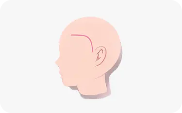
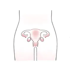
A subarachnoid hemorrhage is a condition in which a bulge in a blood vessel, known as a brain aneurysm, ruptures, causing blood to fill the subarachnoid space (the space between the membranes covering the brain).
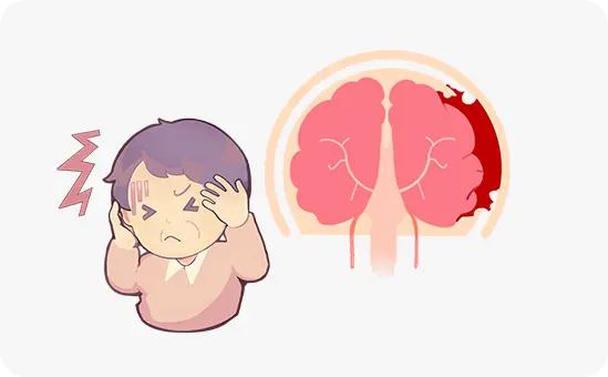
The symptoms of a subarachnoid hemorrhage include intense pain, as if you were hit with a baseball bat at the moment of rupture. Other typical symptoms include nausea and vomiting, impaired consciousness, and visual impairment.
Most subarachnoid hemorrhages occur suddenly without warning. It is a frightening condition that rapidly spreads throughout the brain, causing serious damage. Of the people who develop a subarachnoid hemorrhage, one third return to society, one third will have severe sequelae, and the remaining one third will die.
Treatment of unruptured brain aneurysms
Two methods are available for treating an unruptured brain aneurysm: endovascular treatment (coil embolization) and craniotomy (clipping surgery).
Endovascular treatment (coil embolization)
A metal coil is inserted into the aneurysm to block blood flow and seal it.
-
There are restrictions on the shape and size of the target aneurysm. An aneurysm may recur.
-
Less burden on the body.

Craniotomy (clipping surgery)
A portion of the skull is opened, and a titanium metal clip is used to clip the base of the aneurysm to prevent rupture and bleeding.
-
No restriction on shape; recurrence is extremely rare.
-
Greater burden on the body.
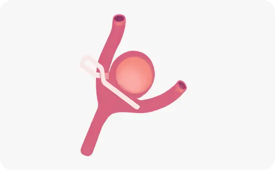
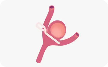
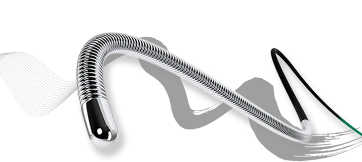
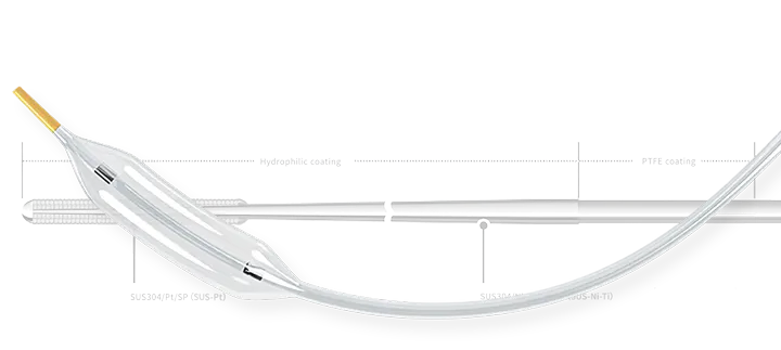
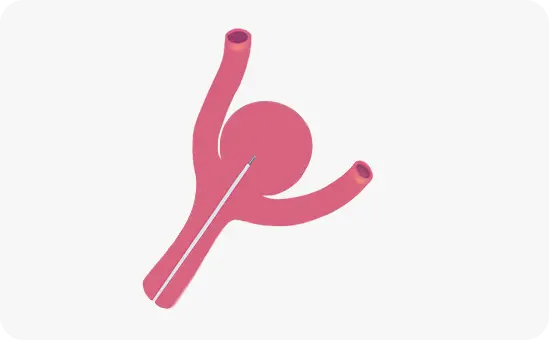
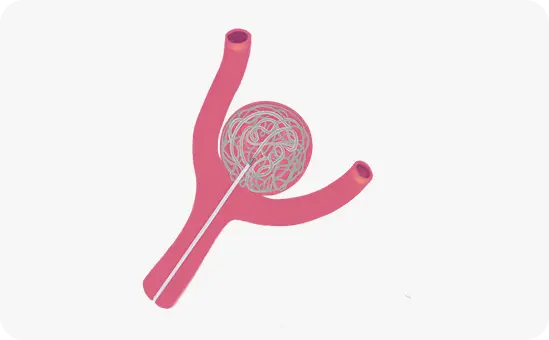
Endovascular treatment: coil embolization
A catheter is inserted into an artery in the wrist or groin. Using this catheter as a guide, a thinner microcatheter is advanced into the aneurysm.
Metal coils are then delivered through the microcatheter and inserted into the aneurysm. The coils fill the aneurysm and seal it, blocking any space for blood flow. This reduces the risk of a rupture.
The coils are not stable if the base of the aneurysm is too wide. To prevent them from slipping out of the aneurysm, a stent may be used in combination with the coils.
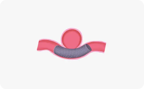
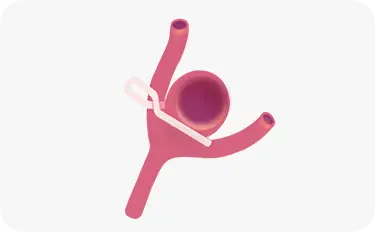
New endovascular treatment: flow diverter stenting
A flow diverter is a stent with a finer mesh than a regular stent. Placing this flow diverter at the base of an aneurysm disrupts the blood flow into the aneurysm. The blood inside the aneurysm slowly turns into a clot, and the aneurysm shrinks or eventually disappears. This treatment does not need to place coils within the aneurysm, so it has a lower risk of rupture during surgery. This treatment is currently used in a limited number of institutions, although its use is expected to become more widespread in the future.
Craniotomy: clipping surgery
A drill is used to remove part of the skull near the temple.
A brain aneurysm often forms in regions deep inside the brain. A surgeon carefully opens the brain folds using a surgical microscope, looking for the aneurysm. Once found, a metal clip is placed across the base of the aneurysm to prevent rupture and bleeding.
Once complete, the surgeon replaces the skull and sutures the muscle and skin.
Clipping surgery is performed under general anesthesia.